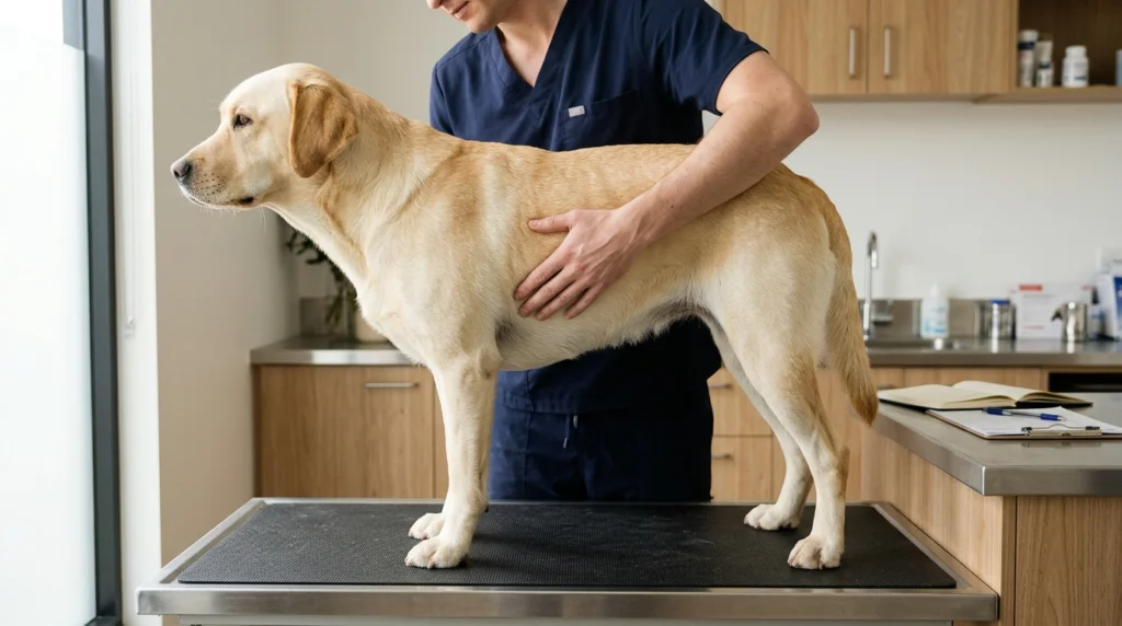
Assess your dog’s anatomical body condition, compute metabolic resting energy requirements (RER), and calculate clinical target feeding portions with safe weight loss or gain timelines.
Dog Profile & Evaluation
Nutritional Adjustment Plan
Weight Loss ProtocolDaily Meal Portions (Copper)
Based on 375 kcal/cupWeigh-in Milestone Schedule
Bi-weekly weigh-in check-in targets| Milestone | Timeline | Target Weight | Progress |
|---|
Explore more companion animal, small pet, and canine care tools in our Interactive Calculators Directory, including our Dog Heat Cycle & Ovulation Window Calculator, Dog Crate Size Calculator, and our Kitten Growth & Feeding Calculator.
Direct Answer Clinical Capsule: Canine Body Condition & Caloric Planning
Important Clinical Notice
Clinical Answer Capsule: The World Small Animal Veterinary Association (WSAVA) and American Animal Hospital Association (AAHA) designate the 9-point Body Condition Score (BCS) system as the clinical gold standard for evaluating canine body composition. An ideal score is BCS 4 to 5 out of 9, corresponding to an optimal body fat percentage of 15% to 20%.
Each integer change above or below 5 represents an approximate 10% to 15% deviation in total body weight. A dog’s true lean target weight is mathematically modeled by:
BWideal = (BWcurrent ÷ [1 + (BCS – 5) × 0.10])
Canine resting energy expenditure scales allometrically via Kleiber’s Law (RER = 70 × BWkg0.75). To reverse canine adiposity without triggering protective muscle catabolism or metabolic slowdown, veterinary guidelines mandate a safe weight reduction velocity of 1.0% to 2.0% of total body weight per week (clinical target: 1.25%/week), utilizing dedicated high-protein, high-fiber therapeutic diets rather than reduced portions of maintenance kibble.
1. The Pathophysiology of Canine Adiposity & Chronic Low-Grade Systemic Inflammation
In contemporary veterinary medicine, canine obesity is no longer classified as a benign cosmetic condition or a simple consequence of caloric imbalance. It is recognized by the World Small Animal Veterinary Association (WSAVA), the American Veterinary Medical Association (AVMA), and the British Small Animal Veterinary Association (BSAVA) as an insidious, progressive, multifactorial chronic neuro-endocrine disease state.
Canine Adiposity Pathophysiology & Systemic Inflammatory Cascade
1. Etiological Trigger: Hypercaloric Intake & Physical Inactivity
2. Cellular Remodeling: White Adipose Tissue (WAT) Hypertrophy & Hyperplasia → Adipocyte Hypoxia & M1 Macrophage Infiltration
3. Endocrine & Cytokine Cascade: Pathological Adipokine Secretion
- Elevated TNF-α & IL-6: Chronic low-grade systemic inflammation
- Hyperleptinemia: Central hypothalamic leptin resistance & continuous polyphagia
- Hypoadiponectinemia: Peripheral insulin resistance & endothelial dysfunction
4. End-Organ Compromise:
- Joint cartilage MMP activation → Accelerated severe osteoarthritis
- Decreased thoracic wall compliance → Pickwickian hypoventilation
- Hepatic lipidosis, systemic dyslipidemia & glomerular hypertension
⚠ Cumulative Clinical Impact: Median Life Span Reduction of 1.8 to 2.5 Years (Kealy et al., 2002)
White Adipose Tissue as an Endocrine Organ
Historically, canine adipose tissue was regarded as an inert depot for triglyceride storage, serving mechanical cushioning and thermal insulation. Landmark molecular discoveries have disproven this passive model: white adipose tissue (WAT) is now understood to be an exceptionally complex, highly vascularized, and metabolically active endocrine and immune organ.
Adipocytes, in concert with stromal vascular fraction cells (pre-adipocytes, endothelial cells, fibroblasts, and resident immune cells), synthesize and release dozens of biologically active signaling peptides collectively termed adipokines, as well as classical pro-inflammatory cytokines:
- Tumor Necrosis Factor-Alpha (TNF-α): Hypertrophied canine adipocytes recruit circulating monocytes that differentiate into classically activated M1 macrophages, forming characteristic “crown-like structures” around dying, hypoxic fat cells. These macrophages secrete excessive TNF-α, which inhibits insulin receptor substrate-1 (IRS-1) phosphorylation in hepatocytes and myocytes, directly driving peripheral insulin resistance. Furthermore, circulating TNF-α induces systemic vascular endothelial dysfunction and upregulates hepatic production of C-reactive protein (CRP).
- Interleukin-6 (IL-6): Approximately one-third of circulating IL-6 originates directly from visceral adipose depots. IL-6 stimulates hepatic acute-phase reactant synthesis (including fibrinogen and serum amyloid A), promoting a pro-thrombotic, hyper-inflammatory state while exacerbating hypothalamic inflammation.
- Leptin: Secreted almost exclusively by mature adipocytes in direct proportion to total fat mass, leptin normally crosses the blood-brain barrier to bind leptin receptors (Ob-Rb) in the hypothalamic arcuate nucleus, stimulating anorexigenic pro-opiomelanocortin (POMC) neurons and inhibiting orexigenic neuropeptide Y (NPY) / agouti-related peptide (AgRP) neurons to induce satiety. In obese dogs, persistent extreme hyperleptinemia downregulates receptor sensitivity, inducing central leptin resistance. The brain perceives a state of intracellular starvation despite massive peripheral energy reserves, compelling the animal to display persistent food-seeking behaviors.
- Adiponectin: In healthy, lean dogs, adiponectin is the most abundant circulating adipokine, exerting potent anti-inflammatory, insulin-sensitizing, and anti-atherogenic actions by activating AMPK and PPAR-α. Crucially, as adipose mass expands, adiponectin gene expression is paradoxically downregulated. Obese dogs suffer from profound hypoadiponectinemia, stripping vascular beds and visceral organs of its protective, anti-fibrotic influence.
Secondary Pathologies & Organ-System Compromise
The biological burden of canine adiposity radiates across every physiological organ system:
1. Accelerated Osteoarthritic Cartilage Degradation
Canine osteoarthritis (OA) was historically viewed as a simple biomechanical “wear-and-tear” phenomenon where excess gravitational load accelerates articular cartilage erosion. While excessive mechanical joint loading is undeniably catastrophic—amplifying ground reaction forces and stress on the cranial cruciate ligaments (CCL), coxofemoral joints, and intervertebral discs—recent research confirms that the inflammatory component of adiposity dominates OA pathogenesis.
Circulating adipokines (TNF-α, IL-1β, and adiponectin dysregulation) directly permeate synovial fluid. Within the joint capsule, these cytokines trigger synovial fibroblasts and chondrocytes to express matrix metalloproteinases (MMPs) and aggrecanases (ADAMTS-4 and ADAMTS-5). These proteolytic enzymes systematically cleave type II collagen and aggrecan, dissolving the extracellular cartilage matrix from within. Consequently, obese dogs develop debilitating joint pathology at younger ages, experience chronic lameness, and require substantially higher doses of non-steroidal anti-inflammatory drugs (NSAIDs), increasing their risk of secondary renal and gastrointestinal toxicity.
2. Cardiorespiratory and Ventilatory Impairment
Adipose deposition does not occur merely subcutaneously; it infiltrates visceral cavities, retroperitoneal spaces, and intercostal musculature:
- Decreased Thoracic Compliance: Massive subcutaneous fat deposits across the ribcage physically restrict thoracic excursion during inhalation. Concurrently, excessive visceral and omental fat pushes the diaphragm cranially into the pleural cavity, compressing functional residual capacity (FRC) and tidal volume.
- Ventilatory Mismatch & Hypoxemia: The obese canine lung operates under constant restrictive load, creating ventilation-perfusion (dot{V}/dot{Q}) mismatches. Dogs exhibit exercise intolerance, exertional dyspnea, and rapid heat retention because panting—the primary canine thermoregulatory mechanism—requires significantly greater mechanical work against an encumbered chest wall.
- Tracheal & Upper Airway Collapse: In toy and small breeds genetically predisposed to tracheal collapse (e.g., Yorkshire Terriers, Pomeranians, Pugs, Chihuahuas), cervical fat deposits exert direct extrinsic compressive pressure on hypocellular, weakened hyaline cartilage rings. In brachycephalic breeds, excessive pharyngeal and soft palate fat accumulation severely narrows the upper airway lumen, transforming mild stenotic breathing into life-threatening Brachycephalic Obstructive Airway Syndrome (BOAS) crises.
3. Metabolic Syndrome and Endocrine Dysfunction
While dogs rarely develop classic human-style type 2 diabetes mellitus (canine diabetes is predominantly an immune-mediated destruction of pancreatic beta cells resembling type 1 diabetes), obese dogs suffer from profound subclinical metabolic syndrome:
- Insulin Resistance & Compensatory Hyperinsulinemia: Peripheral glucose clearance in skeletal muscle is severely impaired. Pancreatic beta cells compensate by secreting copious insulin, creating chronic hyperinsulinemia that fosters vascular smooth muscle hypertrophy, sodium retention, and secondary systemic hypertension.
- Hepatic Steatosis: Chronic non-esterified fatty acid (NEFA) flux from insulin-resistant adipose depots floods the liver, overwhelming beta-oxidation and promoting hepatic lipid accumulation (steatosis), subclinical transaminitis (elevated serum ALP and ALT), and compromised hepatic detoxification.
The 1.8 to 2.5 Year Life Span Reduction (Kealy et al., 2002)
The most conclusive empirical evidence demonstrating the catastrophic impact of canine adiposity is the landmark 14-year Purina Life Span Study led by Dr. Richard D. Kealy and published in the *Journal of the American Veterinary Medical Association* (JAVMA, 2002).
In this rigorously controlled lifelong investigation, 48 paired Labrador retriever littermates were divided into two cohorts:
- Control Group (Ad-Libitum / Overfed): Dogs fed unrestricted maintenance rations, naturally settling at a Body Condition Score of BCS 6.5 to 7.5 out of 9 (representing a typical modern household dog with mild-to-moderate adiposity).
- Restricted Group (Ideal Lean Fed): Littermates fed identical nutrients but restricted by 25% in total caloric energy, maintaining a Body Condition Score of BCS 4.5 to 5.0 out of 9 throughout life.
| Clinical Parameter | Restricted Diet (Ideal BCS 4–5) | Ad-Libitum Diet (Obese BCS 7) |
|---|---|---|
| Median Life Expectancy | 13.0 Years | 11.2 Years |
| Net Difference in Canine Longevity | +1.8 Years (+15%) | -1.8 Years (-15%) |
| Onset of Chronic Osteoarthritis Treatment | 12.0 Years | 9.9 Years (2.1 yrs earlier) |
| Median Age at First Chronic Disease Treatment | 12.0 Years | 9.9 Years |
| Prevalence of Radiographic Hip Dysplasia at Age 2 | 29% | 68% |
| End-of-Life Severe Osteoarthritic Burden | Significantly Reduced & Localized | Severe, Generalized & Disabling |
The clinical findings were profound:
- Median survival was prolonged by 15% (1.8 full years) in the lean-fed cohort. In giant or toy breed scaling, this equates to up to 2.5 human-equivalent decades of vital life.
- Lean-fed dogs delayed the onset of debilitating chronic degenerative diseases—including malignant neoplasia, hip dysplasia, and osteoarthritis—by an average of 2.1 years.
- At 2 years of age, 68% of the ad-libitum fed dogs already exhibited radiographic signs of hip joint osteoarthritis, compared to only 29% of the restricted cohort.
- Maintaining an ideal body condition score is unequivocally the single most effective, clinically proven medical intervention available to extend canine health span and longevity.
2. The 9-Point WSAVA / Purina Body Condition Score (BCS) Matrix
Subjective visual assessment alone is notoriously unreliable; canine coats, deep chests, breed-specific conformations, and human cognitive bias routinely cause owners and clinicians to underestimate canine fat mass. Standardized clinical evaluation requires combining rigorous visual inspection with systematic bimanual palpation using the clinically validated 9-point Body Condition Score matrix developed by Purina and established globally by the World Small Animal Veterinary Association (WSAVA).
Clinical Assessment Protocol
Stand directly over the dog’s spine while they stand square.
Key Finding: Presence, depth, or complete absence of the post-costal hourglass waist constriction.
Position your eyes parallel to the dog’s ventral abdomen at floor level.
Key Finding: Upward abdominal tuck angle from the caudal sternum to the pelvic inlet.
Place flat palms across the lateral rib cage using gentle gliding pressure.
Knuckle Rule: Fist knuckles = BCS 1–3; Back of hand = BCS 4–5; Fleshy base of thumb = BCS 7–9.
Run hands along dorsal lumbar vertebrae, iliac wings, and ischium.
Key Finding: Differentiates sharp protruding bone margins vs. smooth fat blanket vs. thick fat pads.
Complete 9-Point Clinical Staging Matrix
| BCS Tier | Clinical Category | Estimated Body Fat % | Visual Silhouette (Dorsal & Lateral) | Tactile Palpation Findings (Ribs, Spine & Pelvis) | Clinical Action Plan |
|---|---|---|---|---|---|
| BCS 1 | Severely Emaciated | < 5% | Prominent, sharp rib silhouettes; deep, hollow abdominal cavity; no subcutaneous tissue; sharp angular pelvic wings. | Ribs, lumbar spinous processes, and pelvic bones razor-sharp with zero fat padding. Severe muscle atrophy evident. | CRITICAL EMERGENCY: Hospitalization, rule out cachexia/parasitism, initiate stepwise refeeding protocol to prevent refeeding syndrome. |
| BCS 2 | Very Thin | 5% – 9% | Ribs, lumbar vertebrae, and pelvic wings easily visible; dramatic, abrupt abdominal tuck; severe hourglass waist. | Ribs easily palpable with zero intercostal fat cushion. Minimal dorsal muscle tone palpable; pelvic margins prominent. | Comprehensive clinical diagnostic workup (CBC, chemistry, fecal); structured caloric titration at 1.3–1.4 × RER. |
| BCS 3 | Thin | 10% – 14% | Ribs readily visible with minimal lighting angle; pronounced waist tuck behind costal arch; noticeable abdominal incline. | Ribs easily palpated with negligible fat cover; individual dorsal vertebral spines palpable beneath skin. | Caloric adjustment: Increase daily caloric intake by 15%–20%; re-evaluate after 14 days. |
| BCS 4 | Underweight / Lean | 15% – 18% | Ribs easily discernible; distinct, crisp waist tuck from above; proportionate, sharp abdominal tuck in lateral profile. | Ribs easily palpable with a thin, pliable subcutaneous fat layer. Smooth pelvic contours without bony protrusion. | Normal physiological target for certain athletic working dogs and sighthounds. Maintain or minor caloric boost (+5%–10%). |
| BCS 5 | Ideal Weight | 19% – 24% | Ribs not visually prominent but silhouette exhibits clear hourglass waist behind ribs; clean upward abdominal tuck. | Ribs easily palpable under a thin layer of subcutaneous fat (feels like the back of a flat hand); bony prominences smooth. | GOLD STANDARD: Maintain current caloric intake (DER = 1.4–1.6 × RER for neutered adult); continue routine bi-weekly weight logging. |
| BCS 6 | Overweight / Heavy | 25% – 29% | Waist visible from above but rounded and indistinct; lateral abdominal tuck discernible but shallow and diminished. | Ribs palpable with slight finger pressure; discernible slight excess subcutaneous fat layer over ribcage and tail base. | Early intervention window: Restrict daily energy intake by 10%–15%; eliminate high-fat table scraps and treats. |
| BCS 7 | Heavy / Overweight | 30% – 34% | Waist absent from above; dorsal silhouette is straight or rectangular; abdominal tuck noticeably absent or flattened. | Ribs palpable only with deliberate, firm flat-palm pressure; moderate fat cushion over lumbar spine and base of tail. | Veterinary medical intervention: Formulate weight loss plan (1.0 × RERideal); target 1.0%–1.5% weekly weight reduction. |
| BCS 8 | Obese | 35% – 39% | Broad, flat, table-like back; dorsal silhouette bulges convexly outward; abdomen distended with pendulous ventral sag. | Ribs extremely difficult to palpate; requiring deep pressure; heavy fat deposits over lumbar spine, tail base, and neck. | Transition to therapeutic high-protein, high-fiber veterinary metabolic diet; evaluate for concurrent mobility limitations. |
| BCS 9 | Morbidly Obese | ≥ 40% | Massive fat deposits distending lateral trunk; convex abdominal contour; thick fat rolls across neck, thorax, and base of tail. | Ribs impossible to palpate even with heavy force; thick fat pads bury spine and pelvis; fat deposits cover limbs. | Immediate medical management: Full laboratory panel; strict veterinary caloric restriction (0.8–1.0 × RERideal); hydrotherapy. |
The Knuckle Analogy: A Practical Owner-Palpation Calibration Tool
To eliminate ambiguity when educating dog owners during clinical consultations, veterinarians utilize the hand knuckle model:
- BCS 1 to 3 (Underweight): Form your hand into a tight clenched fist. Run the flat pads of your opposite fingers over the protruding knuckles on top of your fist. The sharp, prominent, rigid sensation matches the ribcage of an emaciated or thin dog.
- BCS 4 to 5 (Ideal Body Condition): Lay your hand completely flat on a table with your palm facing down. Run the flat pads of your opposite fingers across the back of your hand over the metacarpal bones. You can readily feel and count each individual bone without pressing deeply, but they feel smooth, rounded, and cushioned by soft skin. This represents ideal canine rib palpation.
- BCS 7 to 9 (Overweight to Morbidly Obese): Turn your hand palm-side up. Press your fingers into the fleshy, muscular mound at the base of your thumb (thenar eminence). Even when pushing firmly, the bone beneath is obscured by a dense, spongy layer of tissue. This mirrors the palpation of a dog whose ribs are buried under pathological subcutaneous adipose blankets.
3. Muscle Condition Score (MCS) vs. Body Condition Score (BCS)
One of the most consequential diagnostic missteps in companion animal medicine is conflating body weight or adipose volume with functional muscular fitness. In 2011, the World Small Animal Veterinary Association (WSAVA) introduced the Muscle Condition Score (MCS) as a mandatory clinical vital sign alongside BCS, body weight, body temperature, pulse, and respiration.
BCS vs. MCS Diagnostic Dissociation Matrix
Evaluates fat deposition and stored metabolic energy reserves.
Anatomical Sites: Rib cage, lumbar waistline, abdominal tuck.
Evaluates active functional lean mass and systemic protein reserves.
Anatomical Sites: Temporalis skull, scapular spine, lumbar vertebrae, wings of ilium.
Crucial Clinical Rule: A dog can be simultaneously BCS 8 (Severely Obese) and MCS Severe Wasting (sarcopenic obesity). Never evaluate body fat and muscle mass as a single composite metric.
The Critical Distinction: Fat Mass vs. Lean Body Mass
- Body Condition Score (BCS): Measures energy reserves stored as subcutaneous, retroperitoneal, and visceral adipose tissue. A dog’s BCS fluctuates dynamically based on positive or negative caloric balance.
- Muscle Condition Score (MCS): Evaluates total skeletal muscle protein stores. Skeletal muscle represents the body’s largest metabolic engine, reservoir of essential amino acids, and primary structural support for arthritic joints.
Crucially, BCS and MCS can vary independently:
- A severely underweight, starved dog (BCS 2/9) may possess normal or only mildly wasted muscle mass if the starvation is acute.
- Conversely, an overtly obese dog (BCS 8/9 or 9/9) can simultaneously suffer from life-threatening, advanced skeletal muscle wasting. This dangerous clinical state is termed Sarcopenic Obesity. In these patients, expansive subcutaneous fat blankets completely mask profound muscular atrophy from visual inspection, emphasizing the non-negotiable requirement for physical palpation.
Sarcopenia vs. Cachexia: Pathophysiological Divergence
Canine Sarcopenia
(Age-Related Senescent Muscle Loss)
- Aging myocytes & progressive peripheral denervation.
- Decline in anabolic hormones (GH, IGF-1, testosterone).
- Slow, insidious muscle protein degradation spanning years.
- Appetite & caloric intake remain intact; BCS often stable (BCS 4–6).
Canine Cachexia
(Disease-Driven Hypercatabolism)
- Underlying systemic pathology: CHF, CKD, neoplasia, sepsis.
- Profound systemic pro-inflammatory cytokine release (TNF-α, IL-1, IL-6).
- Ubiquitin-proteasome hypercatabolism with rapid myofibrillar lysis.
- Rapid, acute muscle breakdown (weeks/months); occurs across ANY BCS (1–9).
Veterinarians differentiate muscle loss into two fundamentally distinct clinical entities:
1. Canine Sarcopenia (Senescent Muscle Loss)
- Etiology: The gradual, age-associated decline in skeletal muscle mass and functional power occurring in healthy geriatric dogs in the absence of primary systemic disease.
- Mechanism: Driven by intrinsic mitochondrial senescence, progressive loss of motor unit innervation (alpha-motor neuron apoptosis), reduced cellular responsiveness to circulating anabolic hormones (insulin-like growth factor-1 [IGF-1] and growth hormone), and mild declines in daily physical activity.
- Nutritional Characteristics: Caloric appetite remains normal; body condition score is frequently preserved at BCS 4 to 6. Sarcopenia responds positively to elevated dietary protein intake (≥ 3.0–3.5 g protein/kg ideal BW), supplemental branched-chain amino acids (BCAAs: leucine, isoleucine, valine), omega-3 fatty acids (EPA and DHA), and low-impact resistance conditioning.
2. Canine Cachexia (Cytokine-Driven Hypercatabolic Muscle Wasting)
- Etiology: Accelerated, systemic muscle wasting secondary to severe chronic diseases, most notably Congestive Heart Failure (CHF) (cardiac cachexia), Chronic Kidney Disease (CKD) (uremic cachexia), and Malignant Neoplasia (cancer cachexia).
- Mechanism: Driven by uncontrolled neuro-hormonal activation and hyper-secretion of catabolic cytokines (TNF-α, IL-1β, IL-6). These inflammatory mediators stimulate the ubiquitin-proteasome pathway within myocytes, systematically dismantling contractile myofibrillar proteins (actin and myosin) to supply hepatic gluconeogenesis and acute-phase protein synthesis.
- Clinical Prognosis: Cachexia cannot be reversed simply by force-feeding additional calories. It signifies an advanced disease state and correlates directly with significantly reduced median survival times.
The 4 Cardinal Palpation Sites for MCS
To grade MCS accurately, clinicians must palpate four distinct anatomical zones where subcutaneous fat deposition is minimal and underlying skeletal muscle contours are directly accessible:
- Temporalis Muscles (Cranial Vault): Stand face-to-face with the dog. Place palms over the parietal bones of the cranium and palpate the muscular convexities over the sagittal crest. Normal muscle feels smooth, full, and convex. In wasting, the sagittal crest of the skull feels sharp, prominent, and framed by deep hollow depressions.
- Scapular Spines (Shoulder Girdle): Place your hand over the lateral aspect of the shoulder blade. Palpate the distinct vertical bony ridge of the scapular spine. In a dog with normal MCS, the supraspinatus and infraspinatus muscles completely fill the fossa, making the bony spine smooth and difficult to isolate. In moderate-to-severe wasting, the scapular spine protrudes like a raised ridge with deep gutters on either side.
- Lumbar Paraspinal Epaxial Muscles: Run your flat palms along either side of the dorsal spinous processes of the lumbar spine (L1–L7). Healthy epaxials feel thick, taut, and cylindrical, projecting upward to or slightly above the vertebral tips. In wasted patients, the epaxials are hollowed and concave, causing the dorsal spines to feel like distinct, sharp pegs.
- Pelvic Wings (Iliac Crests & Ischial Tuberosities): Cup your fingers over the dorsal iliac wings (the hips). In normal musculature, the gluteal muscles round smoothly over the iliac crests. In wasting, the cranial dorsal iliac spines protrude sharply with prominent hollows between the hip bones and tail head.
The 4-Tier WSAVA Muscle Condition Score Classification
| MCS Tier | Clinical Staging | Cranial Temporalis Palpation | Scapular Spine Palpation | Lumbar Epaxial Palpation | Pelvic Wings Palpation |
|---|---|---|---|---|---|
| Normal | Clinically Intact Muscle Mass | Full, convex musculature flush with sagittal crest. No cranial hollows. | Scapular fossa fully filled; bony ridge rounded and masked by muscle. | Epaxials thick, firm, and cylindrical; spine tip barely discernible. | Gluteal mass fully covers iliac crest; smooth, rounded hip contours. |
| Mild Wasting | Early / Subclinical Loss | Flat temporalis profile; slight angular demarcation at sagittal crest. | Scapular spine easily palpable as a distinct ridge, but fossa not hollow. | Epaxial muscles slightly flattened; vertebral tips easily felt. | Iliac wings readily palpable under skin; slight depression in gluteal shelf. |
| Moderate Wasting | Significant Lean Mass Depletion | Definite bilateral hollows above zygomatic arches; sharp sagittal crest. | Scapular spine protrudes sharply; noticeable indentations in fossae. | Epaxial muscles concave; vertebral spines project above muscle line. | Pelvic bones prominent; gluteal muscle mass thin and easily compressed. |
| Severe Wasting | Critical Sarcopenia / Cachexia | Deep cranial depressions; skull appears gaunt, skeletal, and skull-like. | Scapular spine razor-sharp; muscular fossae severely hollowed out. | Severe concave epaxial atrophy; vertebral column feels like peg teeth. | Sharp, angular iliac and ischial bones protruding; near-total loss of gluteal mass. |
4. Mathematical Modeling of Ideal Body Weight
Determining a dog’s ideal body weight (BWideal) is an essential prerequisite for formulating any clinical weight loss or nutritional recovery plan. Estimating target weight through breed-standard averages is inherently flawed due to massive intra-breed genetic variances in frame size, height, and bone density.
Derivation & Mathematical Proof
The mathematical derivation of ideal body weight is founded upon extensive clinical research conducted by Purina, the University of Liverpool Small Animal Weight Management Clinic, and WSAVA nutritionists (Mawby et al., 2004; German et al., 2006).
Empirical dual-energy X-ray absorptiometry (DEXA) body composition scans confirm that:
- BCS 5/9 represents the ideal baseline, where body fat accounts for approximately 15% to 20% of total body mass.
- For each integer unit that a dog scores above BCS 5, the animal carries approximately 10% excess body weight above its lean physiological frame:
- BCS 6 = BWideal × 1.10 (+10% excess mass)
- BCS 7 = BWideal × 1.20 (+20% excess mass)
- BCS 8 = BWideal × 1.30 (+30% excess mass)
- BCS 9 = BWideal × 1.40 (+40% excess mass)
- Conversely, each integer unit below BCS 5 represents an approximate 10% deficit in ideal body mass:
- BCS 4 = BWideal × 0.90 (-10% deficit)
- BCS 3 = BWideal × 0.80 (-20% deficit)
- BCS 2 = BWideal × 0.70 (-30% deficit)
- BCS 1 = BWideal × 0.60 (-40% deficit)
We express current body weight (BWcurrent) as a linear transformation of ideal body weight (BWideal):
BWcurrent = BWideal × [ 1 + (BCS – 5) × 0.10 ]
Solving algebraically for BWideal, we obtain the canonical veterinary formula:
BWideal = (BWcurrent ÷ [1 + (BCS – 5) × 0.10])
Worked Clinical Examples Across Canine Frame Categories
Example 1: Toy Breed — Obese Chihuahua
- Clinical Presentation: Adult spayed female Chihuahua.
- Current Weight (BWcurrent): 4.20 kg (9.26 lb).
- Body Condition Score: BCS = 8 / 9 (Significant adipose blankets, no waist, pendulous abdominal tuck).
- Calculation:
BWideal = (4.20 ÷ [1 + (8 – 5) × 0.10]) = (4.20 ÷ [1 + 3 × 0.10]) = (4.20 ÷ [1.30]) ≈ 3.23 kg (7.12 lb)
- Excess Adipose Mass: 4.20 kg – 3.23 kg = 0.97 kg (2.14 lb).
- Clinical Insight: While 0.97 kg sounds minor to an owner, it represents 23.1% excess pathological body mass, equivalent to an 80 kg human carrying an extra 24 kg of visceral fat.
Example 2: Medium Breed — Overweight French Bulldog
- Clinical Presentation: Adult neutered male French Bulldog.
- Current Weight (BWcurrent): 18.00 kg (39.68 lb).
- Body Condition Score: BCS = 7 / 9 (Loss of waist, rounded trunk, thick neck rolls).
- Calculation:
BWideal = (18.00 ÷ [1 + (7 – 5) × 0.10]) = (18.00 ÷ [1 + 2 × 0.10]) = (18.00 ÷ [1.20]) = 15.00 kg (33.07 lb)
- Excess Adipose Mass: 18.00 kg – 15.00 kg = 3.00 kg (6.61 lb).
- Clinical Insight: In a brachycephalic breed with pre-existing stenotic nares, shedding this 3.0 kg relieves critical mechanical compression off the trachea and soft palate, often resolving nocturnal snoring and exertional stridor.
Example 3: Large Breed — Obese Labrador Retriever
- Clinical Presentation: 6-year-old neutered male Labrador Retriever.
- Current Weight (BWcurrent): 46.00 kg (101.41 lb).
- Body Condition Score: BCS = 8 / 9 (Flat table-back, no abdominal tuck, bilateral hindlimb stiffness).
- Calculation:
BWideal = (46.00 ÷ [1 + (8 – 5) × 0.10]) = (46.00 ÷ [1.30]) ≈ 35.38 kg (78.00 lb)
- Excess Adipose Mass: 46.00 kg – 35.38 kg = 10.62 kg (23.41 lb).
- Clinical Insight: Losing 10.6 kg reduces compressive forces across the stifle joints by up to 40 kg during locomotive push-off, drastically reducing lameness scores.
Example 4: Giant Breed — Morbidly Obese English Mastiff
- Clinical Presentation: 5-year-old spayed female English Mastiff.
- Current Weight (BWcurrent): 88.00 kg (194.01 lb).
- Body Condition Score: BCS = 9 / 9 (Massive subcutaneous fat pads, severe ventral sag, exertional dyspnea).
- Calculation:
BWideal = (88.00 ÷ [1 + (9 – 5) × 0.10]) = (88.00 ÷ [1 + 4 × 0.10]) = (88.00 ÷ [1.40]) ≈ 62.86 kg (138.58 lb)
- Excess Adipose Mass: 88.00 kg – 62.86 kg = 25.14 kg (55.43 lb).
5. Canine Energy Metabolism & Resting Energy Requirements
Calculating daily caloric prescriptions requires a rigorous comprehension of canine bioenergetics. Estimating caloric needs based on crude body weight without physiological scaling produces gross prescription errors that either induce metabolic shock or guarantee weight management failure.
Allometric Metabolic Scaling vs. Linear Approximations
Kleiber’s 0.75 Exponent (RER = 70 × BW0.75) vs. Flawed Linear Model (30 × BW + 70)
| Dog Body Weight | Kleiber Exponential (Accurate) | Linear Formula (30 × BW + 70) | Clinical Error Deviation |
|---|---|---|---|
| 2.0 kg (Toy Chihuahua) | 118 kcal/day | 130 kcal/day | +10.2% (Overfeeds toy dog) |
| 10.0 kg (Beagle) | 394 kcal/day | 370 kcal/day | -6.1% (Acceptable alignment) |
| 30.0 kg (Golden Retriever) | 897 kcal/day | 970 kcal/day | +8.1% (Acceptable alignment) |
| 70.0 kg (Mastiff / Great Dane) | 1,694 kcal/day | 2,170 kcal/day | +28.1% (+476 kcal/day OVERFEED!) |
Kleiber’s Law vs. Flawed Linear Equations
In 1932, Swiss biologist Max Kleiber demonstrated that mammalian basal metabolic rate does not scale linearly with body mass (BW1.0), nor does it scale strictly with Euclidean surface area (BW0.67). Instead, across all homeothermic mammals from a 20-gram mouse to a 4-tonne elephant, basal metabolic rate scales allometrically with body mass raised to the three-quarters power (BW0.75).
The National Research Council (NRC, 2006) and WSAVA designate Kleiber’s Law as the standard formula for canine Resting Energy Requirement (RER):
RER (kcal/day) = 70 × ( BWkg )0.75
In human clinical shorthand, a linear approximation formula is frequently cited in non-specialized literature:
RERlinear (kcal/day) = 30 × ( BWkg ) + 70
Why Linear Approximations are Clinically Dangerous
The linear equation is mathematically tangent to Kleiber’s power curve only between 10 kg and 20 kg. Outside this narrow bracket, the linear formula breaks down catastrophically:
| Canine Body Mass (BWkg) | Kleiber Allometric RER (70 × BW0.75) | Flawed Linear RER (30 × BW + 70) | Absolute Discrepancy (kcal) | Error Magnitude (%) | Clinical Hazard |
|---|---|---|---|---|---|
| 2.0 kg (Toy Chihuahua) | 118 kcal | 130 kcal | +12 kcal | +10.2% | Caloric overfeeding; persistent weight gain in tiny frame. |
| 1.0 kg (Teacup Puppy) | 70 kcal | 100 kcal | +30 kcal | +42.9% | Severe overfeeding; gastrointestinal osmotic overload. |
| 5.0 kg (Yorkshire Terrier) | 234 kcal | 220 kcal | -14 kcal | -6.0% | Underestimation of maintenance energy. |
| 10.0 kg (Beagle / Spaniel) | 394 kcal | 370 kcal | -24 kcal | -6.1% | Minor divergence (acceptable clinical bracket). |
| 20.0 kg (Border Collie) | 662 kcal | 670 kcal | +8 kcal | +1.2% | Convergence zone (minimal error). |
| 40.0 kg (German Shepherd) | 1,113 kcal | 1,270 kcal | +157 kcal | +14.1% | Substantial caloric over-prescription; weight loss stalls. |
| 60.0 kg (Rottweiler / Mastiff) | 1,509 kcal | 1,870 kcal | +361 kcal | +23.9% | Severe caloric surplus; completely defeats weight loss. |
| 80.0 kg (Giant Mastiff) | 1,871 kcal | 2,470 kcal | +599 kcal | +32.0% | Massive surplus (+600 kcal/day = 1.2 kg fat gain per month). |
Clinical Invariant: The linear shortcut must be permanently retired from veterinary protocols. For large and giant breeds, using 30 × BW + 70 prescribes up to 600 excess kcal per day, completely arresting weight loss and baffling owners.
Daily Energy Requirement (DER) Life-Stage Multipliers
Resting Energy Requirement (RER) accounts purely for cellular respiration, thermoregulation, and visceral homeostasis at complete rest in a thermo-neutral room. To determine total Daily Energy Requirement (DER), clinicians apply physiological multipliers reflecting physical activity, reproductive status, and clinical goals:
DER (kcal/day) = Multiplier × RER
| Physiological Status & Clinical Goal | Base Weight Used | WSAVA / NRC Multiplier Range | Recommended Standard Multiplier | Clinical Rationale & Metabolic Dynamics |
|---|---|---|---|---|
| Veterinary Weight Loss (Adipose Reduction) | BWideal | 0.8 to 1.0 × RERideal | 1.0 × RERideal | Forces negative energy balance utilizing ideal frame baseline. For refractory canine obesity, down-titrate to 0.8 × RERideal. |
| Intact Adult (Normal Activity) | BWcurrent | 1.6 to 1.8 × RER | 1.8 × RER | Elevated basal metabolic rate driven by circulating androgens/estrogens. |
| Neutered / Spayed Adult (Normal Activity) | BWcurrent | 1.4 to 1.6 × RER | 1.6 × RER | Loss of gonadal hormones decreases basal metabolic rate by 15%–20%; increased appetite. |
| Sedentary / Prone to Obesity Adult | BWcurrent | 1.2 to 1.4 × RER | 1.3 × RER | Low physical expenditure; prevents insidious adipose accumulation. |
| Senior / Geriatric Canine | BWcurrent | 1.1 to 1.4 × RER | 1.2 × RER | Reduced locomotive activity combined with age-related decline in lean muscle mass. |
| Working Dogs (Light / Moderate Work) | BWcurrent | 1.8 to 2.5 × RER | 2.0 × RER | Herding, assistance, field hunting, or daily active endurance. |
| Working Dogs (Heavy Endurance / Sledding) | BWcurrent | 3.0 to 8.0 × RER | 4.0–5.0 × RER | Extreme muscular exertion in sub-zero environments (e.g., Iditarod canine athletes). |
| Controlled Weight Gain (Underweight BCS 3) | BWcurrent | 1.2 to 1.4 × RER | 1.3 × RER | Controlled positive energy balance without gastrointestinal osmotic distress. |
| Emaciated Refeeding (BCS 1–2 Inpatient) | BWcurrent | 0.25 → 1.0 × RER | Stepwise Titration | Staged caloric escalation over 7–10 days to avert fatal hypophosphatemia. |
6. Veterinary Weight Loss Protocols & Caloric Restriction
Successful canine adipose reduction requires a disciplined therapeutic protocol. Unmonitored or excessively aggressive caloric restriction does not merely fail; it triggers protective biological countermeasures that compromise canine health and ensure rapid rebound weight gain.
The Canine Weight Loss Triad
Target exactly 1.0 × RER of Ideal Body Weight (floor: ≥ 0.8 × RERideal) to drive a safe 1.0% to 1.5% weekly weight loss rate.
Therapeutic high protein (>30% DM), high fiber (>10% DM), and L-carnitine (300–500 ppm) to protect myofibrils and induce satiety.
Underwater treadmill hydrotherapy and low-impact flat-surface leash walking. Strictly zero ballistic jumping or fetch play.
Clinically Validated Rate of Weight Reduction
The American Animal Hospital Association (AAHA) and WSAVA establish the safe velocity of canine weight reduction at:
Safe Rate = 1.0% to 2.0% of Total Body Weight per Week
Optimal Clinical Velocity = 1.25% to 1.50% per Week
Pathological Hazards of Rapid Weight Loss (> 2.0% per Week):
- Severe Sarcopenic Muscle Degradation: Under severe caloric deficits (> 40% restriction), the canine liver exhausts glycogen reserves within 24 hours. The neuro-endocrine axis upregulates hepatic gluconeogenesis, mobilizing systemic amino acids by breaking down functional skeletal muscle. Up to 50% of weight lost during crash dieting consists of vital lean skeletal muscle, devastating physical mobility.
- Adaptive Thermogenesis & Metabolic Slowdown: Rapid weight loss triggers a dramatic down-regulation of circulating triiodothyronine (T3) and basal sympathetic tone. The body enters a state of hypometabolic energy preservation, lowering baseline Resting Energy Requirement by up to 25%. When normal caloric intake resumes, the dog rapidly regains adipose mass—the classic “yo-yo” phenomenon.
- Severe Behavioral Distress & Polyphagia: Extreme caloric deprivation causes uninhibited secretion of ghrelin from gastric oxyntic cells while leptin collapses. Dogs experience persistent, agonizing hunger cues, driving destructive scavenging, pica, aggression around food bowls, and constant vocalization.
Satiety Mechanisms & Dietary Architecture
Achieving long-term weight reduction requires specialized nutritional architecture that manipulates gastrointestinal neuro-endocrine feedback loops:
Therapeutic High-Protein, High-Fiber Diet Metabolic Signaling Pathways
Stimulates duodenal CCK and distal ileal PYY secretion.
→ Central hypothalamic satiety signaling & 100% lean mass preservation
Insoluble fiber promotes gastric stretch; soluble fiber forms a viscous gel delaying gastric transit.
→ Prolonged interprandial satiety & blunted postprandial glucose curve
Upregulates Carnitine Palmitoyltransferase-1 (CPT-1) across mitochondrial membranes.
→ Accelerates mitochondrial β-oxidation of long-chain fatty acids
- High Dietary Protein Concentration (≥ 30% to 35% Dry Matter [DM]):
Elevated dietary protein stimulates duodenal endocrine I-cells to secrete cholecystokinin (CCK) and distal ileal L-cells to release peptide YY (PYY), both of which act directly on the brainstem and hypothalamus to induce satiety. Crucially, high protein availability ensures continuous nitrogen balance, forcing the body to draw exclusively upon adipose triglycerides for energy while sparing skeletal myofibrils.
- Dual-Action Fiber Matrix (≥ 10% to 15% DM Crude Fiber):
- Insoluble Fiber (Purified Cellulose): Adds physical, calorically inert volume to the kibble. This promotes gastric mechanical distension, triggering gastric vagal stretch receptors that signal gastric fullness to the solitary tract nucleus in the brainstem.
- Soluble & Fermentable Fiber (Psyllium, Beet Pulp, Fructooligosaccharides): Absorbs luminal water to form a high-viscosity hydrogel. This slows gastric emptying rates, blunts postprandial glucose and insulin spikes, and fuels colonic colonocytes with short-chain fatty acids (SCFAs: acetate, propionate, butyrate).
- L-Carnitine Supplementation (300 to 500 mg/kg DM):
L-carnitine is an essential cofactor required to transport long-chain fatty acids across the inner mitochondrial membrane via the carnitine palmitoyltransferase-1 (CPT-1) shuttle. Supplementation upregulates mitochondrial beta-oxidation, preserving lean tissue mass while accelerating hepatic fat clearance.
The “Feed Less Maintenance Food” Fallacy
A frequent, disastrous error made by well-intentioned pet owners is attempting weight loss by simply reducing the daily cup measurement of their dog’s regular adult maintenance kibble by 30% to 50%.
⚠ The Micronutrient Malnutrition Trap: Why Cutting Maintenance Kibble Fails
Commercial adult maintenance foods are balanced on a per-calorie basis assuming normal maintenance DER. When an owner reduces daily maintenance rations by 35%:
- Total Protein Intake: Deficient by 35% → Direct trigger for sarcopenic skeletal muscle catabolism.
- Trace Minerals (Zinc, Selenium, Iron): Deficient by 35% → Impairs thyroid deiodinase and immune defense.
- Essential Micronutrients (Choline, B-Vitamins, Calcium): Below minimum physiological thresholds.
- Clinical Consequence: Rebound polyphagia, coat alopecia, lethargy, and immediate weight rebound.
Clinical Solution: Switch to a purpose-formulated veterinary therapeutic reduction diet with nutrient-to-calorie fortification.
Every complete and balanced commercial dog food is formulated to meet AAFCO Nutrient Profiles or FEDIAF Guidelines on a per-calorie basis assuming normal maintenance intake (DER). When an owner reduces daily maintenance rations by 35%:
- Micronutrient Starvation: The dog receives 35% less calcium, phosphorus, zinc, copper, selenium, B-complex vitamins, and vitamin E than the physiological minimum required for basal cellular maintenance. Over 12 to 24 weeks, dogs exhibit dry, brittle hair coats, epidermal scaling, impaired wound healing, and subclinical immune suppression.
- Accelerated Sarcopenia: Standard maintenance diets contain only 20% to 24% crude protein. Under caloric restriction, this marginal protein concentration is inadequate to prevent negative nitrogen balance, accelerating muscle wasting.
- Clinical Directive: Canine weight loss must always be conducted using a dedicated veterinary therapeutic weight loss diet (e.g., Hill’s Prescription Diet r/d or Metabolic, Royal Canin Satiety Support, Purina Pro Plan Veterinary Diets OM). These formulas are engineered with nutrient-to-calorie fortifications, delivering 100% of essential micronutrients and elevated protein within a severely reduced caloric footprint.
7. Weight Gain & Safe Refeeding Protocols for Malnourished/Rescue Dogs
While managing canine adiposity dominates urban small animal practice, rehabilitating emaciated, starved dogs (BCS 1/9 to 2/9) poses an acute, life-threatening metabolic challenge. Rescuers and owners instinctively want to offer large bowls of rich food to a starving dog; in veterinary critical care, uncontrolled refeeding is frequently fatal.
⚠ Life-Threatening Refeeding Syndrome Pathophysiology Cascade (BCS 1–2)
Phase 1: Starvation State → Glycogen depletion, reliance on fat/ketones, total-body depletion of intracellular PO4, K+, and Mg2+.
Phase 2: Insulin Surge → Rapid glucose uptake triggers massive insulin spike, driving remaining PO4, K+, and Mg2+ into cells for glycolysis.
Phase 3: Severe Extracellular Depletion → Acute severe hypophosphatemia (< 1.5 mg/dL), acute hypokalemia, and hypomagnesemia.
Phase 4: Fatal Multi-System Decompensation:
- Erythrocyte ATP depletion → Acute hemolytic anemia
- 2,3-DPG depletion → Tissue hypoxia & diaphragmatic respiratory failure
- Hypokalemia / Hypomagnesemia → Lethal cardiac arrhythmias, tetany & seizures
⛔ Outcome if Unchecked: Cardiorespiratory Collapse within 24 to 72 Hours
The Pathophysiology of Refeeding Syndrome
During prolonged starvation:
- Glycogen stores in hepatocytes and skeletal myocytes are completely exhausted within 24 to 36 hours.
- The body transitions its entire bioenergetic machinery to fatty acid beta-oxidation, ketone body utilization, and proteolysis.
- Crucially, total body intracellular stores of electrolytes—most notably inorganic phosphorus (P), potassium (K^+), and magnesium (Mg2+)—become severely depleted, although serum concentrations may initially appear normal on blood panels due to extracellular fluid contraction and dehydration.
When an emaciated dog is abruptly fed a large, carbohydrate-dense meal:
- The rapid influx of luminal glucose triggers an immediate, massive surge of insulin from pancreatic beta cells.
- Insulin instantly activates cellular sodium-potassium ATPase pumps and glucose transport proteins, driving glucose, potassium, magnesium, and phosphate out of the extracellular serum and into the intracellular compartment to fuel glycolysis and phosphorylation.
- Acute, Profound Hypophosphatemia (< 1.5 mg/dL): Phosphate is an essential substrate for synthesizing adenosine triphosphate (ATP) and 2,3-diphosphoglycerate (2,3-DPG). Without phosphate:
- Acute Hemolytic Anemia: Erythrocytes lose membrane deformability and structural integrity, undergoing catastrophic intravascular hemolysis.
- Diaphragmatic Paralysis: Respiratory muscles fail due to bioenergetic exhaustion, resulting in progressive hypoventilation and fatal hypoxia.
- Cardiomyopathic Collapse: Myocardial contractility plummets, triggering refractory ventricular arrhythmias, muscle fasciculations, seizures, and death within 24 to 72 hours of refeeding.
Staged Refeeding Protocol for Emaciated Canines (BCS 1–2)
Veterinary guidelines mandate a conservative, stepwise caloric reintroduction over a 10-day timeline:
| Staging Day | Caloric Prescription | Daily Meal Frequency | Clinical Monitoring & Goals |
|---|---|---|---|
| Days 1 to 2 | 25% of Current RER | 4–6 Micro-Meals (q4h) | Low-carb formulation; establish baseline serum phosphorus, K+, Mg2+. |
| Days 3 to 4 | 50% of Current RER | 4–5 Small Meals | Recheck electrolyte gate. If PO4 drops >0.5 mg/dL, halt advancement. |
| Days 5 to 6 | 75% of Current RER | 3–4 Small Meals | Gradual 25% daily blend transition to recovery target diet. |
| Days 7 to 9 | 100% of Current RER | 3 Meals | Euvolemia, normophosphatemia, and normal gut transit confirmed. |
| Day 10+ | 1.2 to 1.4 × Target RER | 2–3 Standard Meals | Steady anabolism; safe weekly weight gain of 1.0% to 2.0%. |
| Staging Day | Caloric Prescription (% of Baseline RER) | Daily Meal Frequency | Dietary Composition Guidelines | Clinical Monitoring & Laboratory Gates |
|---|---|---|---|---|
| Days 1 to 2 | 25% of Current RER | 4 to 6 Micro-Meals (every 4 hours) | Highly digestible, low-carbohydrate, moderate-to-high fat, high-quality animal protein diet (e.g., veterinary recovery liquid or canned critical care diet). | Check baseline serum electrolytes (P, K, Mg), PCV/TS, and blood glucose every 12 to 24 hours. Supplement potassium and phosphate parenterally if serum levels trend downward. |
| Days 3 to 4 | 50% of Current RER | 4 to 5 Small Meals | Maintain identical low-carbohydrate nutritional density. Warm meals to body temperature (38.5^circC) to enhance palatability and reduce thermoregulatory burden. | Palpate abdomen for gastric distension, ileus, or vomiting. Re-verify serum phosphorus is > 2.5 mg/dL. |
| Days 5 to 6 | 75% of Current RER | 3 to 4 Meals | Begin gradual 25% incorporation of the long-term recovery puppy/performance diet. | Monitor body weight. Ensure zero dependent edema or tachypnea (signs of volume overload). |
| Days 7 to 9 | 100% of Current RER | 3 Meals | 100% target diet transition. Caloric intake now matches basal maintenance at resting state. | Electrolytes should be fully stabilized. Stools should be formed and firm. |
| Day 10+ | 1.2 to 1.4 × RERtarget | 2 to 3 Meals | Controlled caloric surplus designed for steady tissue accretion. | Target weekly weight gain of 1.0% to 2.0% of total body weight per week. Re-evaluate BCS and MCS every 14 days. |
8. Multi-Modal Physical Conditioning & Exercise Prescription
Nutritional restriction achieves caloric deficit, but physical conditioning determines tissue partition kinetics—ensuring the body mobilizes adipose triglycerides while maintaining or building skeletal myofibrils. In canine patients with elevated Body Condition Scores (BCS 7–9), prescribing exercise requires meticulous biomechanical risk mitigation.
High-Impact Exercise Contraindications in Obese Dogs
Owners frequently attempt to accelerate canine weight loss by throwing tennis balls, running long distances on asphalt, or encouraging jumping. In an overweight or obese dog, these activities are strictly contraindicated:
- Joint Ground Reaction Forces (GRF): High-velocity deceleration, cutting, and jumping exert ground reaction forces exceeding 2.5 to 4.0 times body weight across the stifle and carpal joints. In an already compromised, inflamed joint, this mechanical overload precipitates complete rupture of the cranial cruciate ligament (CCL), meniscal tears, and acute exacerbation of subchondral bone remodeling.
- Thermoregulatory Heatstroke Risk: Canine subcutaneous fat blankets act as potent thermal insulators, while visceral fat restricts diaphragmatic movement and respiratory panting efficiency. Obese dogs undergoing high-impact exercise in moderate ambient temperatures (> 20^circC / 68^circF) face extreme risks of exertional hyperthermia, heatstroke, and fatal disseminated intravascular coagulation (DIC).
Hydrotherapy & Underwater Treadmill (UWTM) Biomechanics
Veterinary physical rehabilitation relies on aquatic therapy as the gold standard modality for obese and arthritic canines. The physics of water immersion uniquely resolves the conflict between exercise expenditure and joint preservation:
Moderate gravitational reduction. Excellent for conditioning and building concentric limb propulsion.
Near-weightless environment. Eliminates joint concussion for dogs with severe hip/stifle osteoarthritis or CCL tears.
- Buoyancy & Joint Decompression (Archimedes’ Principle): When a dog is submerged in water to the level of the greater trochanter (hip), buoyant forces offload approximately 62% of the dog’s gravitational body weight. Stifle-height water offloads approximately 38% of body weight. This near-weightless environment eliminates articular concussion, allowing dogs with severe hip dysplasia or stifle osteoarthritis to exercise vigorously without pain.
- Hydrostatic Pressure: The pressure exerted by fluid molecules against submerged limbs stimulates lymphatic and venous return, dramatically reducing periarticular soft tissue edema and joint effusion.
- Viscous Drag & Resistance Conditioning: Water provides viscous resistance that is hundreds of times greater than air. Every step taken on an underwater treadmill requires concentric muscle contraction across the quadriceps, biceps femoris, and gluteal muscle groups, actively building functional lean body mass (reversing sarcopenia) while burning calories.
- Clinical UWTM Protocol: Begin with 2 to 3 sessions per week. Initial sessions utilize water filled to the greater trochanter at 28^circC to 30^circC (82^circF to 86^circF). Treadmill speed is set to a comfortable walking pace (1.0 to 1.5 mph) for 3 to 4 walking intervals of 2 to 3 minutes, separated by 1-minute rest periods. Walk intervals are increased by 10% weekly as cardiovascular stamina improves.
Controlled Flat-Surface Leash Walking Protocol
For pet owners without access to veterinary rehabilitation facilities, structured leash walking represents the foundational conditioning pillar:
| Conditioning Stage | Walk Duration | Daily Frequency | Terrain & Surface Protocol |
|---|---|---|---|
| Weeks 1 to 2 | 10–15 Minutes | 2x Daily | Flat turf or forgiving level pavement; steady cadence. |
| Weeks 3 to 4 | 15–20 Minutes | 2x Daily | Flat surfaces; introduce gentle 5% natural inclines. |
| Weeks 5 to 6 | 20–25 Minutes | 2x Daily | Mixed level park terrain; continuous brisk walking pace. |
| Weeks 7 to 8+ | 25–30 Minutes | 2x Daily | Varied surfaces; sustained aerobic conditioning. |
- Environmental Control: Walks must be conducted on flat, forgiving surfaces (turf, dirt trails, or level asphalt). Loose gravel, deep sand, and steep downhill descents must be avoided, as downhill locomotion transfers up to 70% of ground reaction forces onto forelimb carpal and elbow joints.
- Harness Mechanics: Dogs must be walked in a properly fitted, padded Y-shaped chest harness that does not restrict scapular translation or compress the cervical trachea. Choke chains, pinch collars, or neck collars that press against the ventral trachea are contraindicated, especially in brachycephalic or toy breeds.
- The “Rule of 10%”: Walking duration should increase by no more than 10% per week. If a dog demonstrates post-exercise stiffness, hesitation to rise, or behavioral reluctance to walk the following morning, duration must be immediately de-escalated to the previous week’s baseline for 7 days.
9. Breed Conformation Caveats & False Diagnoses
Applying standardized body condition scoring algorithms without accounting for breed morphology is a primary cause of misdiagnosis in veterinary practice. Morphological divergence across the domestic dog (*Canis lupus familiaris*) exceeds that of any other terrestrial vertebrate species. Clinicians and owners must calibrate their tactile and visual criteria against four distinct conformational phenotypes:
Deep keel, extreme abdominal tuck, physiological body fat 8–12%.
Palpation Rule: Gauge temporalis and gluteals; last 2 ribs are naturally visible.
Barrel chest, flat costal arch; cylindrical rib cage conceals fat deposits.
Palpation Rule: Palpate 5th–7th ribs and tailhead; waistline is naturally absent.
Dense woolly undercoat completely masks skeletal contours and adiposity.
Palpation Rule: Visual check useless; firm bimanual compression beneath coat required.
Elongated axial skeleton with extreme Hansen Type I IVDD disc vulnerability.
Palpation Rule: Zero tolerance for BCS > 5; even minor weight gain accelerates spinal paralysis.
1. Dolichocephalic Sighthounds (Greyhounds, Whippets, Salukis)
Sighthounds possess anatomical specializations selected over millennia for high-speed cursorial hunting:
- Physiological Adipose Baseline: Fit adult sighthounds normally carry a body fat percentage of 8% to 12%, compared to 15% to 20% in standard mesomorphic breeds.
- Anatomical Norms: An aerodynamic, deep thoracic keel with an extreme, natural abdominal tuck is physiological. In a healthy, athletic Greyhound or Whippet, the outlines of the last two to three ribs and dorsal tips of the lumbar spinous processes should be faintly visible in normal standing posture.
- Common False Diagnoses: Well-intentioned non-specialist clinicians and shelter personnel frequently misdiagnose fit sighthounds as “emaciated” (BCS 2 or 3) and advise owners to fatten them up. Excess weight forces these light-boned athletes into BCS 6+, predisposing them to catastrophic joint failure and metabolic strain.
- True Pathological Indicators: In a sighthound, true malnutrition is identified by:
- Deep temporal muscle hollows on the cranium (MCS wasting).
- Prominent, sharp protrusion of the tuber coxae (hip points) accompanied by hollowed gluteal fossae.
- Palpable loss of cervical and shoulder muscle mass.
2. Brachycephalic & Barrel-Chested Breeds (Bulldogs, Pugs, Boston Terriers)
In brachycephalic, stocky breeds, conformational geometry obscures standard landmarks:
- The “Substance and Bone” Myth: Kennel club standards historically praise “cobby,” “heavy,” and “thick-set” builds. Owners and breed judges frequently mistake morbid obesity (BCS 8/9) for desirable “breed substance.”
- Absent Hourglass Waist: The extreme cylindrical curvature of the ribcage in English Bulldogs and Pugs means they rarely exhibit a pronounced dorsal hourglass indentation, even when at an ideal body fat percentage.
- Tactile Diagnostic Anchor: Clinicians must ignore the visual waist and rely strictly on flat-palm compression over the 5th through 7th ribs. If the ribs cannot be felt without forceful thumb gouging, the dog is overweight.
- BOAS Escalation: In brachycephalic dogs, excess body fat is concentrated heavily in the soft palate, tongue base, and pharyngeal tissues, drastically narrowing the already compromised upper respiratory tract and accelerating Brachycephalic Obstructive Airway Syndrome (BOAS).
3. Heavy Double-Coated Breeds (Newfoundlands, Malamutes, Golden Retrievers)
Dense, double-layered hair coats (consisting of a short, woolly undercoat beneath long, coarse guard hairs) completely distort visual assessment:
- The Visual Optical Illusion: A heavy double coat adds 2 to 4 inches of visual circumference around the chest and flank, making a lean dog appear stocky or an obese dog appear massive.
- The Neglect Hazard: Conversely, in severely neglected dogs with heavy matting or pelting, a thick coat can completely conceal life-threatening cachexia and emaciation (BCS 1/9). A dog can starve to death under a thick coat without the owner recognizing muscle wasting.
- Mandatory Bimanual Palpation: Visual scoring is clinically invalid in these breeds. The clinician must part the fur with both hands and firmly press flat palms through the coat directly onto the costal arch, dorsal lumbar vertebrae, and pelvic wings.
4. Chondrodystrophic Breeds & Intervertebral Disc Disease (Dachshunds, Corgis)
In breeds with hereditary chondrodysplasia (characterized by FGF4 retrotransposon insertions on chromosome 12, causing premature degeneration and calcification of the nucleus pulposus):
- Mechanical Axial Load: An elongated vertebral column supported by short legs creates an extraordinary mechanical bending moment across the thoracolumbar junction (T11–L2).
- The IVDD Trigger: Each 0.5 kg of excess adipose tissue dramatically increases compressive and shear forces across degenerating discs. Excess body weight transforms manageable, low-grade disc protrusions into catastrophic Hansen Type I intervertebral disc herniations (IVDD), resulting in acute spinal cord compression, loss of deep pain sensation, and irreversible paraplegia.
- Clinical Directive: Chondrodystrophic breeds must be maintained at a strict BCS 4.0 to 4.5 out of 9 (lean body condition). There is zero clinical tolerance for “mild” weight gain in a Dachshund.
10. 12-Question Comprehensive Clinical FAQ
Q1: What is the difference between the 5-point and 9-point canine BCS systems?
Clinical Answer: The 9-point Body Condition Score (BCS) system developed by Purina and adopted by the World Small Animal Veterinary Association (WSAVA) provides twice the diagnostic granularity of the older 5-point scale. On the 9-point scale, each integer point deviation above or below ideal (BCS 5) correlates linearly to approximately 10% to 15% change in body fat mass. The 5-point scale lacks sensitivity to detect early 10% adipose shifts, allowing subclinical obesity to progress unmanaged. Veterinary consensus universally favors the 9-point scale for clinical practice and clinical nutritional management.
Q2: How does canine obesity reduce life expectancy by nearly two years?
Clinical Answer: The landmark 14-year Purina Life Span Study (Kealy et al., 2002) conducted on Labrador retrievers proved that dogs maintained at an ideal body condition (BCS 4.5 to 5.0) lived a median of 1.8 to 2.5 years longer (13.0 years vs. 11.2 years) compared to ad-libitum fed littermates (BCS 6.5 to 7.5). White adipose tissue functions as a metabolically active endocrine organ that continuously secretes pro-inflammatory cytokines (TNF-alpha, IL-6) and adipokines. This chronic systemic inflammation accelerates the onset and severity of osteoarthritis, induces insulin resistance, impairs cardiopulmonary performance, and significantly hastens the development of chronic degenerative diseases.
Q3: What is the mathematical formula used to calculate a dog’s ideal body weight?
Clinical Answer: A dog’s ideal body weight (BWideal) is calculated using the clinically validated inverse linear formula:
BWideal = (BWcurrent ÷ [1 + (BCS – 5) × 0.10])
Because each unit above BCS 5 represents approximately 10% excess body weight (e.g., BCS 7 corresponds to 20% overweight, or 1.20 times ideal weight; BCS 9 corresponds to 40% excess weight, or 1.40 times ideal weight), dividing current weight by this factor accurately isolates the dog’s lean target frame.
Q4: Why is Kleiber’s exponential law (70 * BW^0.75) superior to linear RER formulas (30 * BW + 70)?
Clinical Answer: Kleiber’s Law (RER = 70 × BWkg0.75) is grounded in allometric metabolic scaling, reflecting the physiological reality that metabolic rate is proportional to surface area and metabolic body size rather than simple linear mass. The linear approximation equation (RER = 30 × BWkg + 70) is only mathematically accurate for dogs between 10 kg and 20 kg. For toy breeds under 5 kg, linear formulas substantially underestimate resting caloric needs, creating hypoglycemia risk; for large and giant breeds over 30 kg, linear equations dramatically overestimate caloric requirements, causing weight loss failure.
Q5: What is the maximum safe weekly rate of weight loss for an overweight dog?
Clinical Answer: The clinically validated safe rate of canine weight reduction is 1.0% to 2.0% of total body weight per week, with an optimal clinical target of 1.25% to 1.5% per week. Restricting calories too severely to exceed 2.0% weekly loss causes undesirable loss of lean skeletal muscle mass, triggers metabolic slowdown through adaptive thermogenesis, induces profound behavioral food obsession, and risks hepatic lipid accumulation.
Q6: Why can’t I just feed smaller portions of my dog’s regular adult kibble for weight loss?
Clinical Answer: Standard over-the-counter adult maintenance diets are formulated to meet AAFCO nutrient profiles only when fed at normal maintenance caloric intakes. Restricting maintenance food by 25% to 40% induces proportionate deficiencies in essential amino acids, calcium, phosphorus, trace minerals (zinc, selenium), and water- and fat-soluble vitamins. Furthermore, maintenance foods lack the elevated protein-to-calorie and fiber-to-calorie ratios required to preserve lean muscle mass and maintain gastric distension satiety, leading to sarcopenia and chronic hunger vocalization.
Q7: What is the difference between Muscle Condition Score (MCS) and Body Condition Score (BCS)?
Clinical Answer: BCS evaluates adipose tissue (body fat) distribution across the ribcage, waist, and abdominal profile. MCS specifically evaluates lean skeletal muscle mass over four distinct bony prominences: the cranium (temporalis), scapulae (shoulder blades), thoracic/lumbar vertebrae, and pelvic wings (iliac crests). A dog can be morbidly obese (BCS 9/9) while simultaneously suffering from severe muscle wasting (sarcopenic obesity) due to chronic kidney disease, congestive heart failure, or hyperadrenocorticism.
Q8: What is Refeeding Syndrome, and why is it fatal in emaciated dogs (BCS 1-2)?
Clinical Answer: Refeeding Syndrome is a life-threatening neuro-metabolic complication that occurs when an emaciated, starved animal abruptly receives large quantities of carbohydrate-rich food. During starvation, cellular electrolytes (phosphate, potassium, magnesium) are depleted, and energy relies on ketone and fat catabolism. Rapid nutrient reintroduction triggers an immediate insulin surge, which forces phosphate and potassium from extracellular serum into cells for glycolysis and ATP synthesis. The resulting acute hypophosphatemia causes erythrocyte hemolysis, respiratory failure, cardiac arrhythmias, seizures, and death within 24 to 72 hours.
Q9: How should an emaciated rescue dog (BCS 1 to 2) be safely refed?
Clinical Answer: Severely underweight dogs must be refed using a conservative stepwise protocol under veterinary supervision. Day 1 starts at 25% of current Resting Energy Requirement (RER) divided into 4 to 6 small, low-carbohydrate, high-fat, moderate-protein meals. If serum phosphorus and electrolytes remain stable, intake increases to 50% on Days 2-3, 75% on Days 4-5, and 100% of RER by Day 7. Only after 10 to 14 days of metabolic stability should caloric intake be gradually escalated above maintenance (1.2 to 1.4 × RER) to support gradual tissue accretion.
Q10: Why do visual BCS assessments fail on heavy double-coated dog breeds?
Clinical Answer: Dense, plush double coats (such as those of Newfoundlands, Great Pyrenees, Chow Chows, and Alaskan Malamutes) conceal the lateral abdominal tuck and dorsal hourglass waist silhouette, creating the visual illusion of obesity. Conversely, in heavily neglected or matted dogs, a thick coat can mask life-threatening cachexia and emaciation. Veterinary guidelines mandate hands-on bimanual palpation beneath the coat, using firm flat palms across the ribs, spine, and pelvis.
Q11: How should Body Condition Score be adjusted for naturally lean sighthounds?
Clinical Answer: Sighthound breeds (such as Greyhounds, Whippets, Salukis, and Italian Greyhounds) possess distinct physiological adaptations: a higher percentage of lean muscle mass, an aerodynamic deep thoracic keel with an extreme natural abdominal tuck, and lower baseline body fat percentages (often 8% to 12% in peak fitness compared to 15% to 20% in standard mesomorphic breeds). In fit sighthounds, the outlines of the last 2 to 3 ribs and dorsal vertebral tips are normally visible. However, visible hip bones (iliac wings) or sunken temporalis muscles remain pathological indicators of true malnutrition.
Q12: What type of exercise is safe for dogs with a Body Condition Score of 8 or 9?
Clinical Answer: High-impact activities (such as chasing balls, running, jumping into cars, or agility) are strictly contraindicated for severely obese dogs due to extreme mechanical forces on compromised stifle joints (cranial cruciate ligaments), hip joints, and intervertebral discs, as well as risks of heat stroke and cardiorespiratory collapse. Prescribed exercise must be strictly low-impact: hydrotherapy or underwater treadmill walking (where water buoyancy supports 60% to 80% of body weight), followed by controlled, flat-surface leash walking for 10 to 15 minutes twice daily, gradually progressing as body mass decreases.
11. Real-World Clinical Case Studies
Case Study 1: Canine Metabolic Rehabilitation — Morbidly Obese Labrador Retriever
- Signalment: “Buster”, 7-year-old neutered male Labrador Retriever.
- Initial Presentation: Buster presented with severe bilateral pelvic limb lameness, difficulty rising from recumbency, and exertional dyspnea. He was receiving daily non-steroidal anti-inflammatory therapy (deracoxib, 1.5 mg/kg PO q24h) with declining efficacy.
- Baseline Clinical Metrics:
- Body Weight: 48.5 kg (106.9 lb)
- Body Condition Score: BCS 9 / 9 (Morbidly Obese)
- Muscle Condition Score: Mild Wasting (Lumbar epaxial flattening under dense adipose pads)
- Radiographs: Severe bilateral coxofemoral osteoarthritis with prominent periarticular osteophytosis.
- Nutritional & Mathematical Derivation:
BWideal = (48.5 ÷ [1 + (9 – 5) × 0.10]) = (48.5 ÷ [1.40]) = 34.64 kg (76.37 lb)
RERideal = 70 × (34.64)0.75 = 70 × 14.28 = 999.6 ≈ 1,000 kcal/day
DERtarget = 1.0 × RERideal = 1,000 kcal/day
- Therapeutic Intervention:
- Dietary Transition: Abruptly ceased table scraps and adult maintenance kibble. Transitioned over 7 days to a veterinary therapeutic metabolic diet (2,980 kcal/kg, 34% crude protein, 12% crude fiber, supplemented with 400 mg/kg L-carnitine). Daily allotment: 335 grams/day divided into 2 strictly measured meals using a digital gram scale.
- Exercise Protocol: High-impact fetch was strictly prohibited. Enrolled in underwater treadmill hydrotherapy (2×weekly, water at hip height) alongside controlled flat-surface leash walks starting at 10 minutes 2×daily.
- 28-Week Clinical Outcome:
- Weekly Rate of Weight Loss: Averaged 1.15% of total BW per week (0.50 to 0.55 kg/week).
- End-of-Protocol Weight (Week 28): 34.8 kg (Net loss of 13.7 kg / 30.2 lb).
- BCS & MCS: Improved from BCS 9/9 to BCS 5/9 (Ideal); MCS improved from Mild Wasting to Normal (hypertrophy of quadriceps and gluteals from UWTM).
- Mobility Resolution: Deracoxib NSAID therapy was successfully tapered and fully discontinued at Week 20. Buster demonstrated spontaneous trot locomotion, climbed stairs without assistance, and exhibited zero exertional stridor.
Case Study 2: Emaciated Rescue Pointer Nutritional Resuscitation
- Signalment: “Bella”, 2-year-old female intact English Pointer, rescued from severe physical confinement and neglect.
- Initial Presentation: Presented in lateral recumbency, hypothermic (36.8^circC / 98.2^circF), bradycardic (52 bpm), with pale mucous membranes (CRT > 2.5 sec).
- Baseline Clinical Metrics:
- Body Weight: 12.8 kg (28.2 lb)
- Body Condition Score: BCS 1 / 9 (Severely Emaciated)
- Muscle Condition Score: Severe Wasting (Profound temporal hollows, razor-sharp scapular spines, concave epaxials)
- Estimated Breed-Standard Ideal Frame: 21.5 kg (47.4 lb)
- Serum Biochemistry: Serum Phosphorus: 2.1 mg/dL (Markedly Low, ref: 2.5–6.0 mg/dL); Albumin: 1.9 g/dL; PCV: 24%.
- Critical Care Refeeding Protocol:
- Baseline RER Calculation:
RERcurrent = 70 × (12.8)0.75 = 70 × 6.75 = 472.5 kcal/day
- Day 1–2 (25% RER): Prescribed 118 kcal/day delivered as 6 micro-feedings of 20 kcal every 4 hours using a veterinary liquid convalescence diet (low carbohydrate, high branched-chain amino acids). Oral potassium phosphate supplementation initiated (0.5 mmol/kg/day).
- Day 3–4 (50% RER): Serum phosphorus stabilized at 2.8 mg/dL. Caloric intake advanced to 236 kcal/day in 5 micro-feedings. Body temperature normalized to 38.4^circC.
- Day 5–6 (75% RER): Advanced to 354 kcal/day across 4 feedings. Bella was ambulatory and wagging tail. Zero peripheral edema.
- Day 7–9 (100% RER): Reached baseline resting requirement of 473 kcal/day across 3 meals. Transitioned 50% to high-protein puppy growth kibble.
- Day 10 to Week 16 (Anabolic Weight Gain Phase): Escalated to 1.3 × RER of current weight, recalibrated weekly.
- 16-Week Clinical Outcome:
- Weekly Rate of Gain: Steady, controlled average gain of 1.35% per week (0.45 to 0.55 kg/week).
- End-of-Protocol Weight (Week 16): 21.2 kg (46.7 lb).
- BCS & MCS: Improved from BCS 1/9 to BCS 5/9 (Ideal); MCS recovered fully from Severe Wasting to Normal (complete filling of temporalis and gluteal fossae). Serum albumin normalized to 3.3 g/dL; PCV normalized to 42%. Zero episodes of refeeding syndrome or hemolytic anemia occurred.
12. Verified Peer-Reviewed Scientific Bibliography
- WSAVA Global Nutrition Committee. (2021). *WSAVA Nutritional Assessment Guidelines: Body Condition Score & Muscle Condition Score Systems*. World Small Animal Veterinary Association, Global Nutrition Committee Recommendations. Available via WSAVA Consensus Documents.
- National Research Council (NRC) Nutrient Requirements of Dogs and Cats. (2006). *Nutrient Requirements of Dogs and Cats*. The National Academies Press, Washington, DC. ISBN: 978-0-309-08678-3. [Canonical reference for Kleiber allometric energy scaling, basal resting metabolism, and amino acid minimums].
- Kealy, R. D., Lawler, D. F., Ballam, J. M., Mantz, S. L., Biery, D. N., Greeley, E. H., Lust, G., Segre, M., Smith, G. K., & Stowe, H. D. (2002). *Effects of diet restriction on life span and age-related changes in dogs*. Journal of the American Veterinary Medical Association (JAVMA), 220(9), 1315–1320. DOI: 10.2460/javma.2002.220.1315.
- Brooks, D., Churchill, J., Fein, K., Linder, D., Michel, K. E., Tudor, K., Ward, E., & Witzel, A. (2014). *2014 AAHA Weight Management Guidelines for Dogs and Cats*. Journal of the American Animal Hospital Association (JAAHA), 50(1), 1–11. DOI: 10.5326/JAAHA-MS-6331.
- German, A. J. (2006). *The growing problem of obesity in dogs and cats*. The Journal of Nutrition, 136(7), 1940S–1946S. DOI: 10.1093/jn/136.7.1940S.
- German, A. J., Holden, S. L., Bissot, T., Hackett, R. M., & Biourge, V. (2010). *Dietary energy restriction and successful weight loss in obese client-owned dogs*. Journal of Veterinary Internal Medicine, 21(6), 1174–1180. DOI: 10.1111/j.1939-1676.2007.tb01934.x.
- Lund, E. M., Armstrong, P. J., Kirk, C. A., & Klausner, J. S. (2006). *Prevalence and risk factors for obesity in adult dogs from private US veterinary practices*. International Journal of Applied Research in Veterinary Medicine, 4(2), 177–186.
- Kleiber, M. (1932). *Body size and metabolism*. Hilgardia: A Journal of Agricultural Science, 6(11), 315–353. DOI: 10.3733/hilg.v06n11p315.
- Mawby, D. I., Bartges, J. W., d’Avignon, A., Laflamme, D. P., Moyers, T. D., & Cottrell, T. (2004). *Comparison of various methods for estimating body fat in dogs*. Journal of the American Animal Hospital Association (JAAHA), 40(2), 109–114. DOI: 10.5326/0400109.
- Freeman, L. M. (2012). *Cachexia and sarcopenia: Emerging syndromes of importance in dogs and cats*. Journal of Veterinary Internal Medicine, 26(1), 3–17. DOI: 10.1111/j.1939-1676.2011.00838.x.
- Michel, K. E., Anderson, W., Kubanis, P., & Sorenmo, K. U. (2011). *Evaluation of a muscle condition scoring system for assessing body composition in dogs*. American Journal of Veterinary Research, 72(6), 770–777. DOI: 10.2460/ajvr.72.6.770.
- Laflamme, D. P. (1997). *Development and validation of a body condition score system for dogs*. Canine Practice, 22(4), 10–15. [Original validation study of the Purina 9-point BCS matrix against DEXA body fat scans].